The grass spiders (family Agelenidae) are common spiders throughout North America and Europe, and the genus Hololena is a group endemic to the Western U.S. (Chamberlin and Ivie, 1942). As members of this group have been used in studies of toxins and silks, it is of interest to develop a systematic treatment of them which documents their morphology. Such data can be helpful in developing a phylogenetic hypothesis of the relationships between the species in a group, which is something that has not yet been attempted for this genus. This project is an attempt to document some of the morphology of a member of this group using Scanning Electron Microscopy, as many of the interesting morphological characters are difficult, if not impossible, to deal with practically using other imaging techniques.
The common local species of this genus is Hololena adnexa. It builds webs in grasses, trees, and shrubbery. It can be roughly identified by its flat web with a funnel off to one side, where the spider hides. This funnel is often hidden within a leaf, a curled piece of bark, or even a hole in a post or some manmade item. The spider is yellowish-gray, 12-20 mm long (including legs) with two black "racing stripes" on the dorsal side of the abdomen.
The specimen to be imaged was collected from a shrub on campus, euthanized by cooling in a -20°C freezer, and dissected into two parts for fixation-- the anterior end of the cephalothorax, including the head and first two pairs of legs, and the posterior end, including the 3rd and 4th leg pairs and the abdomen. These parts were then fixed in 2% glutaraldehyde for 2 hours, rinsed 3 times in sodium cacodylate buffer (15 min. per rinse), and post-fixed in 1% osmium tetroxide in sodium cacodylate buffer overnight.
Following fixation, the specimen was dehydrated via alcohol series (10 min each @ 35, 50, 70, 80, 90, 95, 100, and 100% ethanol), then critical-point dried. In future preparations, since spiders are normally stored in 70 or 90 percent ethanol, and their soft tissues that require glutaraldehyde and osmium fixation are of less interest for spider systematics, some of the fixation steps could be foregone, starting the drying series at whatever concentration the animal was stored in. However, any imaging of soft tissues, such as silk glands, would require thorough fixation. Carbon dots were used to mount specimens onto stubs, and these could be then immediately sputter-coated, since the carbon dots do not require drying to prevent outgassing into the sputter-coater, which requires a vacuum to function properly.
Before sputter-coating, the specimens were further dissected so that the individual legs and parts of the abdomen and thorax could be imaged individually. The sputter-coating process, once the chamber was pumped down to the proper near-vacuum pressure, took approximately 2.5 minutes to reach a thickness of 20-25 nanometers. Scanning in the SEM then proceeded on several of the sample stubs.
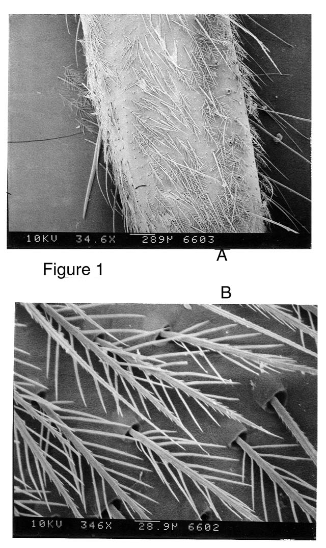
Figure 1, A-B. Femur (large upper leg segment) of leg 4. The objects of interest here are the "cuticular scales"-- the fine feathery scales covering large portions of the spider's cuticle. These would be categorized as "plumose" due to their feather-like morphology (Towsend and Felgenhauer, 1998). Their function is unknown.
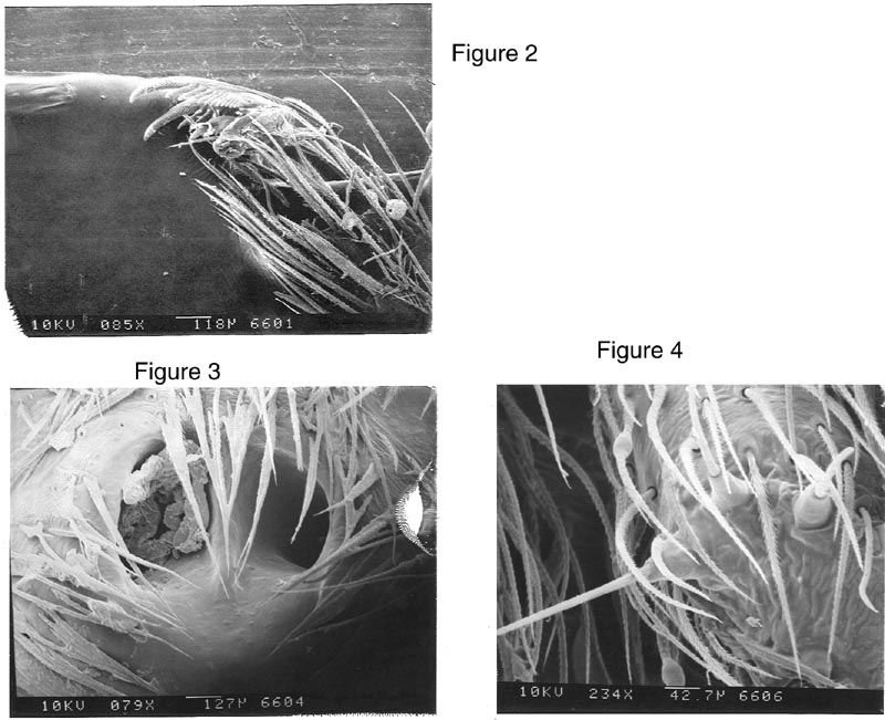
Figure 2. Tarsal claws, leg 4. These claws allow the spider to grab onto and travel about its web. The number of claw teeth (the comblike scales on the bottom of the curved structure) is potentially useful as a taxonomic character.
Figure 3. Epigynum. This is the external genitalic structure of the female. Its shape, and the ridge between the two openings, as well as features out-of-frame relative to this scan are used to determine the species when somatic (body) characters are too similar. The "plug" in the left opening is most likely the sperm-plug of a previous arachnid suitor.
Figure 4. Posterior median spinneret. This spinneret creates silk used to reinforce the dragline that the spider trails behind it, or drops on. Three silk-spigots can be seen extending from the end of the PMS.
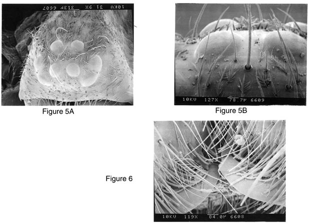
Figure 5. A. Head of spider, including eye-pattern. Eye numbers and patterns are extremely important in the classification of spiders, as is relative size and distance between eyes. This spider has two "procurved" (forward curving) rows of 4 eyes each, with the lateral eyes closer together than the median eyes. This spider also has a zig-zag row of large hairs that can be seen proceeding from between the ALE's (Anterior Lateral Eyes- the large hair in fig 5 B.) back down the dorsum of the cephalothorax. These may serve a sensory purpose.
Figure 6. Close-up of the chelicerae (jaws) and fangs. Looking carefully below and between the hairs hanging down over the chelicerae, cheliceral teeth (seen as triangular projections pointing toward the up-curved fangs) can be seen. The number, size, location, and position of these teeth are all used in spider classification.
This attempt to image various portions of the grass spider has yielded some preliminary taxonomic information as well as some insights into how best to image these animals in the future. Interesting and unexpected was the almost complete coverage of the animal with plumose cuticular hairs. However, hairs were also a problem in some cases, obscuring the epigynum and cheliceral teeth. It would be interesting to attempt to purposely "clear" some of these large hairs from the areas on the surface that require detailed analysis. There are two reasons to believe this might work-- the first in that in some scans, such as fig 1A, it is clear that some hairs have been "rubbed off" and that on sockets remain where the hairs inserted. The second is the overall brittleness of the critical-point dried specimens. It seems reasonable that some sort of "clearing" could be done to better visualize the sclerotized regions which are of greatest taxonomic interest.